BlastoSPIM Models

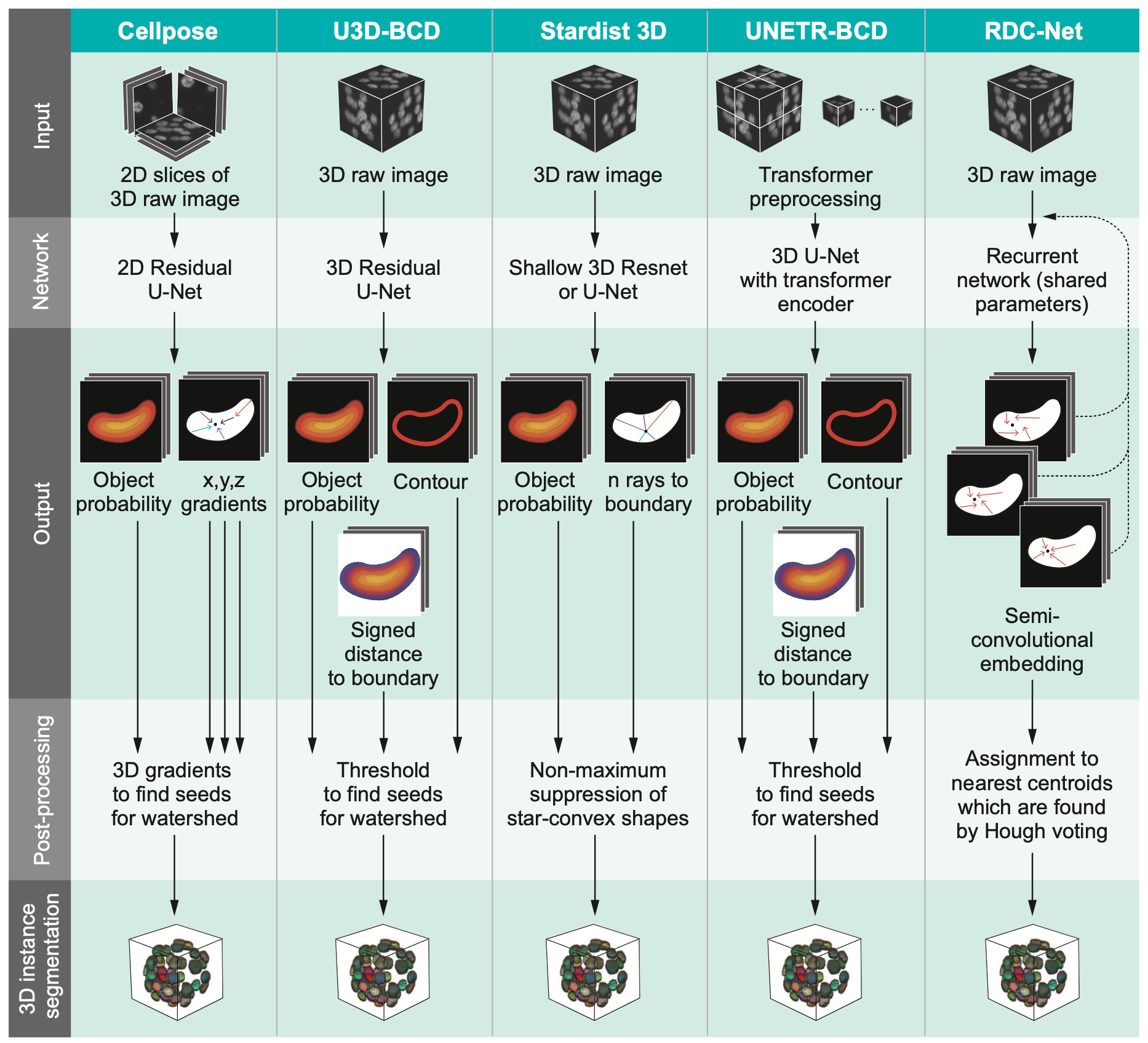
Comparison of five deep-learning-based methods for nuclear instance segmentation

In the illustration to the left each column depicts a different method. From top to bottom, the rows illustrate how images are inputted into the network, the network’s architecture, network outputs for a single nucleus, the post-processing steps, and the 3D instance segmentation, respectively.

Click the image for a detailed view.

Qualitative evaluation of five instance-segmentation networks trained on BlastoSPIM 1.0 and tested on a 62-cell embryo

Instance contours overlaid on a representative slice of the intensity image in xy (top panels) and in xz (bottom panels). Each panel is labelled as: Ground-truth, Stardist for Stardist-3D results, and similarly for other methods. Grey arrows indicate false negatives, including undersegmentation. White arrows denote false positives. Scale bars: 20 μm. Note that false positives and false negatives are defined by comparing the 3D instance segmentation results rather than the results shown in a single 2D slice

Click the image for a detailed view.

Model comparison table.

On BlastoSPIM 1.0, we trained and evaluated five neural network architectures to determine which method achieves the most accurate instance segmentation on our images of preimplantation mouse embryos. The results of the evaluation are summarized in the table to the right.

The model with the best performance was 3D Stardist https://arxiv.org/abs/1908.03636.

Click the image for a detailed view.

Performance results on moderate SNR Images per developmental stage for both the Stardist-3D early embryo model and the Stardist-3D late blastocyst model. This test set includes test images from both BlastoSPIM 1.0 and 2.0. Model hyperparameters were fixed for both models across all stages. The model trained on BlastoSPIM 2.0 starts to outperform the model trained on BlastoSPIM 1.0 during the transition between the 32-cell stage and the 64-cell stage.

Click the image for a detailed view.

Use the links on the right to download two Stardist models optimized for different stages of embryonic development.

Please consult the README files of the archives for model training details.

Contact

  • Address

    Flatiron Institute
    162 Fifth Avenue
    New York, NY 10010
    United States
  • Email

    eposfai at princeton.edu
    lbrown at flatironinstitute.org
    hnunley at flatironinstitute.org